


Clinical examination - venous ulcers
In many cases, a look at the skin surrounding the ulcer can be enough to help you with making the diagnosis " venous ulcer." These patients usually have signs of advanced venous insufficiency - for example, brownish pigmentation called hemosiderin. Simplified, this is rust in the superficial layers of the skin. When there is venous insufficiency, the pressure in the veins increases, and some blood cells manage to leak out of the veins into the surrounding tissue. Red blood cells contain hemoglobin which contains iron. These iron deposits turn brown over time. When you see hemosiderin, you can be quite sure that the patient has venous insufficiency. Note, however, that some other conditions like previous trauma to the leg, chronic edema of different etiology, diabetes, hypertension, and other cardiovascular disease can cause similar iron deposits in the skin.

Figure 1 Hemosiderin deposits in both legs in a patient with venous insufficiency. Typically you will see these pigment changes on the shins and inside areas of the lower legs. Venous insufficiency is the most common cause of these iron deposits in the skin but be aware that other conditions also can lead to this, i.e., hemosiderin is not exclusive to venous insufficiency.
Many patients with chronic venous insufficiency, which has been ongoing for many years, will develop thickened and hard subcutaneous tissue. This is a condition called lipodermatosclerosis. This usually appears in the same areas where we also see hemosiderin deposits. This is because lipodermatosclerosis is also caused by blood vessels leaking - but here, it is the proteins that leak out with blood that cause these changes in the subcutaneous fat. The subcutaneous layers lose their elasticity, and blood circulation is impaired locally, which also predisposes to the development of ulcers.
The changes due to lipodermatosclerosis are often most prominent in the lower third of the legs. This can lead to the characteristic " champagne bottle appearance" in these patients where the lower third of the legs is hard and narrow. In contrast, the upper part of the legs are wide. When you see "champagne bottle" shaped legs in a patient, you can be sure that the venous insufficiency is quite severe and has been ongoing for some time. Note that off-the-shelf medical compression stockings often will not fit these patients, and they often require individually tailored compression garments. In the African healthcare setting, this will be a challenge to acquire.

Figure 3 "Champagne bottle " appearance in the legs of a patient with advanced chronic venous insufficiency.
After several years of chronic venous insufficiency, some patients develop pale/white patches of thin skin called atrophia blanche ( French for " atrophic white"). These changes often appear on the inside of the ankle areas but can appear anywhere on the legs - mainly in the lower thirds. Patients with atrophia blanche changes have to be followed more closely - they are more likely to develop venous ulcers in the future. If an ulcer has developed in an area of atrophia blanche, it will be surprisingly hard to heal even with adequate compression. In our experience, venous ulcers in atrophia blanche areas take 2-3 times longer to heal than other venous ulcers!

Figure 4 Relatively big patches of atrophia blanche behind the medial malleolus of the left leg. Note also some areas of hemosiderin around these areas. ofstore områder med atrofia blanche rundt mediale malleol. Legg også merke til hemosiderin avleiringer i huden omkring malleolen.

Figure 5 Right foot with atrophia blanche around the heel area where small ulcers already have developed. Never underestimate the seriousness of these small ulcerations - they can quickly get out of control and increase in size! This patient has to start with compression immediately! Note that the skin is reddish due to chronic inflammation- this is not an infection, and the patient does not need antibiotics. Note also the hemosiderin deposits farther up the leg.
When the tissue in the legs is swollen over more extended periods, it can become inflamed, and the skin becomes reddish. This is called venous dermatitis or stasis dermatitis. The skin can develop changes that look like eczema. Many misinterpret the red color of the skin as an infection, and not rarely are these patients given antibiotics. There is no indication for antibiotics here- these patients need compression treatment. Topical steroid creams ( Category III-IV steroids creams for 7-10 days) are also beneficial ( in addition to compression treatment). In cases with mild dermatitis, zinc paste or zinc impregnated dressings can be used ( in addition to compression treatment).
The dermatitis condition reduces the skin's immune defense, and these patients are more prone to getting secondary fungal infections. If you see the skin condition worsening despite adequate compression and maybe even steroid creams, you should suspect a secondary fungal infection. Some steroid creams contain antimicrobial agents- for example, Betnovate with chinoform. You must also remember that sometimes venous dermatitis may be a bacterial infection.

Figure 6 A case with venous dermatitis in an early fase. The skin is only slightly reddish and flaky. This patient did not use compression previously.

Figure 7 Venous dermatitis in both legs. When you see redness in both legs, it is improbable that the reason for the redness is caused by a bacterial infection! There is no need for antibiotics here. This patient needs compression treatment and preferably topical steroid cream applied daily for best effect. Note the shape of the legs - they have a slight champagne bottle appearance which indicates that there probably is lipodermatosclerosis in the lower parts of the legs.

Figure 8 A patient with advanced venous insufficiency with chronic venous dermatitis and lipodermatosclerosis. Note the tight mark on the lower third of the leg. The compression bandage was applied wrongly and stopped at the lower third of the leg.
When we see these classical signs: hemosiderin, lipodermatosclerosis, atrophia blanche, and venous dermatitis, we know that the venous insufficiency is more severe. In these cases, we must inform the patient of the importance of life-long compression treatment.

Figure 9 Varicose veins are a sign of early venous insufficiency. However, they alone are usually insufficient to spontaneously give rise to venous ulcers. Often the ulcers develop from some form of trauma to the leg or due to blisters developing in severe edema. The patient should be informed about the need to use compression treatment daily to prevent the venous insufficiency from advancing too rapidly. Surgical removal of varicose veins is indicated when the patient has a venous ulcer or recurrent venous dermatitis. Surgical removal for cosmetic reasons is the most common reason patients opt for surgery.
Ankle-brachial index (ABI)
Ideally, everyone treating venous leg ulcers should have access to measuring the ABI. This is not realistic in most healthcare settings in Africa but rest assured that many health workers in Europe or the United States don't have access to this either.
Why is it so important to measure the ABI? For one, you need it as a diagnostic tool. Is it really a venous ulcer we are dealing with, or could it actually be an arterial ulcer that mimics the appearance of a venous ulcer? Is it maybe a mixed ulcer - both venous and arterial? If the ABI is < 8, an arterial component is involved.
Another reason for measuring the ABI is determining what intensity of compression the patient will tolerate. If you measure an ABI <8, this tells you that you have to choose a milder form of compression, and with an ABI < 5, you should not use compression at all ( unless you are an expert and can monitor the patient very closely. If you plan to use a compression bandage, we recommend a 2-layer short elastic bandage. These are easier to apply and can remain on the leg for up to one week.
If the ABI is < 8, you should use a milder version of these short elastic bandages. Most producers have coined the milder versions as "lite" as in "light as opposed to heavy" ( like Coca Cola light versus the regular Coca Cola). 3M, for example, sells these types of bandages under the name Coban and Coban LITE. The lite versions can be used down to an ABI of 0,5. As you will be aware, it is advantageous to be able to measure the ABI, and if your workplace has the resources, you should push the administration to invest in a handheld doppler.
We have written a separate chapter on doing an ABI measurement properly. Go to "menu" - "practical skills" - "ABI" to read more.
Ultrasound with colour doppler
In most countries, venous ulcers are usually treated in primary health care. If an ulcer does not improve within six weeks, the patient should be referred to a wound care clinic or other higher-level health care provider. There, more diagnostics have to be done to find out why the ulcer does not want to heal.
One of the tests that should be done is an ultrasound, preferably with colour doppler ( duplex) capabilities.
At some centres, this is performed by vascular surgeons; at other clinics, this is done by radiologists. With an ultrasound examination, the severity of the venous insufficiency can be mapped, and leakage in perforator veins can be clearly visualised. Perforator veins are communicating veins between the deep and the superficial venous systems. If such a defective perforator vein is located right beneath a venous ulcer, the wound may never heal without surgical interventions where the perforator vein is closed by different methods. An ultrasound can also detect if the patient has a blockage of the deep veins due to, for example, a previous deep venous thrombosis.

Figure 10 An ultrasound examination with color doppler is a valuable method to find out why a venous ulcer does not heal despite adequate compression therapy.
Blood tests
Laboratory tests are usually not necessary for venous ulcers. However, blood tests may be relevant if a venous ulcer does not show signs of improvement within six weeks after starting compression treatment. Usually, only routine blood tests are necessary ( Hemoglobin, HbA1c, liver- and kidney tests, and electrolytes). These tests may uncover an underlying condition that can impair wound healing. The most obvious example is anemia - if the hemoglobin is lower than 8g/dl, many wounds do not heal or, at best, very slowly. Liver- and kidney diseases can also have a negative effect on wound healing.
Bacterial swabs
There is no need to take bacterial swabs from venous ulcers routinely. It is entirely normal to find lower concentrations of pathological bacteria in all venous ulcers ( and in all other chronic wounds). If you do a bacterial swab from a venous ulcer, you will most certainly find Staphylococcus aureus and probably also enterococcal species in the wound bed.
Only do a bacterial swab if you suspect an infection requiring antibiotics treatment. Then you need to know which bacteria colonize the ulcer to optimize the choice of antibiotic. Remember that bacterial swabs from chronic wounds should always be done after wound debridement/wound cleansing to ensure that we "catch" the bacteria attached to the wound bed.
Sometimes we do a bacterial swab from wounds even though we don't see signs of infection. This is usually when an ulcer does not heal despite other adequate measures. In some cases, bacteria like streptococci and pseudomonas aeroginosa can colonize the wound so that they impair wound healing without causing apparent signs of infection.

Figure 11 E As a rule of thumb: Only do a bacterial swab if you suspect infection and plan to start with antibiotics. Remember that all chronic wounds contain some pathogenic bacteria, but this does not mean that they represent a problem or that they impair wound healing.
Biopsy
If a venous ulcer does not improve within six weeks from the start of compression, you may have to reconsider your wound diagnosis. Could it be a malignant ulcer? Especially squamous cell carcinoma and basal cell carcinoma can mimic venous leg ulcers.
Therefore, it is recommended internationally to do a biopsy from the wound if the wound is not responding to adequate treatment. Biopsies are usually done in local anesthesia with, for example, 1% lidocaine.
A punch biopsy from 3-6 mm is useful for taking such biopsies. Take several biopsies, usually at least three from the wound edges and one from the center of the ulcer. These are placed in formalin as a transport medium on the way to the laboratory. The three biopsies can be sent in one transport vial.
Read more on how to take a biopsy from chronic wounds under the menu "practical skills."
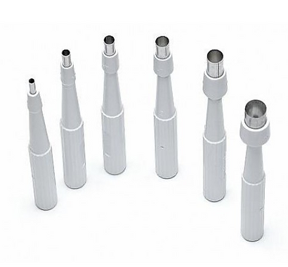
Figure 11 Biopsy punches are available in a range of sizes. The larger the diameter of the biopsy punches, the easier it is for the pathologist to find something relevant. However, a large diameter punch will also leave a large hole that may bleed somewhat. We usually use punches from 3-6mm in diameter in wound care.