


Calciphylaxis skin ulcerations
Treatment of calciphylaxis wounds
Pain management in calciphylaxis
Patients with severe chronic kidney failure, and especially those receiving dialysis, are prone to developing chronic wounds. They are also more likely to develop malignant wounds. A special type of ulceration that can develop in patients with serious kidney disease is calciphylaxis. Of course, not every wound that occurs in patients with chronic kidney failure is a calciphylaxis wound. However, if mysterious, painful necroses develop, this diagnosis should be suspected promptly, and the patient should be referred to specialist healthcare services for multidisciplinary treatment.
Calciphylaxis represents one of the most serious dermatological complications of chronic kidney disease. The prognosis is poor, with a reported one-year mortality of 50–80%, where sepsis from secondarily infected wounds is the most common cause of death. Early recognition and rapid initiation of treatment are therefore crucial.
General
Calciphylaxis is also called calcifying uremic arteriolopathy. It is caused by calcification of small arteries in the skin and adipose tissue. The condition most often manifests in the skin and subcutaneous tissue as painful necrosis, but in rare cases it may also affect other organs.
The condition is most often seen in overweight women with advanced renal failure. Most patients who develop calciphylaxis are therefore dialysis patients. Among dialysis patients, the prevalence is estimated at 1–4%, whereas the incidence in the general population is significantly lower. However, it is important to be aware that it can also occur in patients with normal kidney function or in those with mild renal failure.
Because these patients often have significant comorbidities, they have a reduced immune defense and a higher risk of secondary infection. This can then develop into a life-threatening condition. They often respond less well to antibiotic treatment and therefore have a greater risk of sepsis. With large necroses, the toxic effects of the necrotic tissue itself may worsen kidney function.
The typical clinical picture is that patients develop skin necrosis (often with a black eschar) that eventually progresses into open wounds that fail to heal. Most often, calciphylaxis wounds develop on the abdomen or the lower extremities, but they may occur anywhere on the body. The wounds are almost always extremely painful.
In many cases, the patient has recently started warfarin (Marevan) and sometimes corticosteroids before the necrosis developed. The combination of kidney failure together with these medications therefore gives a higher risk of calciphylaxis necroses.
Five findings that should make you suspect calciphylaxis
1. Extreme pain
Pain that is clearly stronger than the skin findings would suggest is often an early sign.
2. Patient with kidney failure or on dialysis
Calciphylaxis most often occurs in patients with advanced chronic kidney disease.
3. Mottled or purple skin changes that gradually become indurated and painful
4. Rapid development of necrosis
Skin lesions can progress from plaques to necrotic wounds over days to weeks.
5. Location in fat-rich areas
Typical locations include:
-
thighs
-
abdomen
-
buttocks

Figure 1. Calciphylaxis wounds often begin as reddish-purple spots that gradually become darker. They may initially resemble bruises after minor trauma, but an important diagnostic sign is that these skin changes are often very painful in calciphylaxis. Over time, they develop into deeper necroses with even more severe pain.
Copyright: Niels Olson; https://creativecommons.org/licenses/by-sa/3.0/
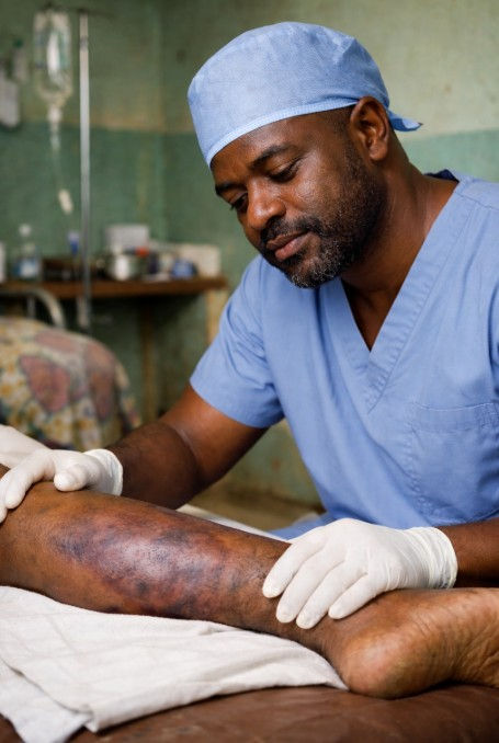
Figure 1a. In pigmented skin, the early purple discoloration that often precedes skin ulcerations in calciphylaxis may be less obvious. Again, a telltale sign that something more serious may be going on is the pain out of proprtion for what you would expect.
Why does calciphylaxis occur?
The pathogenesis of calciphylaxis is complex and not fully understood. The central mechanism is acute calcification of small arterioles in the dermis and subcutaneous fat tissue. In patients with chronic kidney disease, disturbances in mineral metabolism often occur, contributing to calcium deposition in the vessel wall.
Calcification of the vessel wall leads to:
-
increased vascular stiffness
-
endothelial injury
-
microthrombosis (small blood clots)
-
The result is reduced blood supply to the skin and subcutaneous tissue, leading to ischemia and, eventually, tissue necrosis.
Warfarin (Marevan) is also believed to play a role in the pathogenesis. The drug inhibits vitamin K–dependent proteins, which normally help protect against vascular calcification.

Figure 2: Calciphylaxis wounds do not always begin with the classic black skin necrosis—sometimes only reddish ulcerations are seen, as shown here.
Copyright: angiologist.com (as of 2026, this website was no longer online for unknown reasons).

Figure 3: Classic calciphylaxis necrosis on the back of the lower leg with black skin necrosis that developed rapidly over a few days. The patient has renal failure (on dialysis) and had recently started both warfarin and corticosteroids before the necroses appeared. This patient case is described by Dr. Caroline Fife in her blog. We would like to recommend her website, where she has collected many interesting cases involving challenging wounds. Click on the image to visit her website.
Copyright: Dr. Caroline Fife; https://carolinefifemd.com/2018/06/06/dont-miss-this-calciphylaxis-calcific-uremic-arteriopathy/

Figure 4: Calciphylaxis wounds often develop on the abdomen or the lower legs, but they may occur anywhere on the body — including the feet or toes. The yellow-brown skin discoloration is often seen in patients with advanced renal failure.
Risk factors for developing calciphylaxis
Strongly associated risk factors
-
End-stage renal disease
-
Dialysis treatment
-
Hyperphosphatemia
-
Secondary hyperparathyroidism
-
High calcium–phosphate product
-
Warfarin therapy
These factors are closely linked to disturbances in mineral metabolism and vascular calcification.
Moderately associated risk factors
-
Diabetes mellitus
-
Obesity
-
Female sex
-
Hypoalbuminemia
-
Use of calcium-based phosphate binders
-
High doses of vitamin D
Possible risk factors
(reported in case reports or smaller studies)
-
Systemic corticosteroid therapy
-
Liver disease
-
Malignancy
-
Autoimmune diseases
-
Protein C or protein S deficiency
These factors may contribute to an increased tendency toward thrombosis or metabolic disturbances.
Diagnosis
The diagnosis of calciphylaxis is primarily clinical, based on characteristic skin changes in a patient with relevant risk factors.
1. Laboratory tests should include:
-
calcium
-
phosphate
-
parathyroid hormone (PTH)
-
albumin
-
inflammatory markers
2. Imaging
Imaging, such as a plain X-ray of an extremity, may show small calcifications beneath the skin, but these changes can also occur in patients with renal failure without calciphylaxis. Therefore, radiological examinations are often not very helpful for diagnosis.
3. Skin biopsy
A skin biopsy is often not specific, but in some cases, it may support the diagnosis by demonstrating:
-
calcification of small arterioles
-
thrombosis
-
fat tissue necrosis
However, a biopsy should be carefully considered, as the procedure may worsen skin lesions or lead to the development of new ulcerations at the biopsy sites.
Differential diagnoses
Calciphylaxis can be difficult to diagnose because the condition can resemble several other diseases.
Important differential diagnoses include:
-
necrotizing fasciitis
-
vasculitis
-
pyoderma gangrenosum
-
warfarin-induced skin necrosis
-
cholesterol embolization
-
critical peripheral ischemia
Therefore, a thorough clinical assessment and evaluation of the patient’s risk profile are essential.
Treatment of calciphylaxis wounds
There is still some disagreement about the best way to treat calciphylaxis wounds. In most cases, surgical debridement is required as well as systemic therapy. The approach should be relatively aggressive, removing all necrotic tissue until healthy, bleeding tissue is reached. This procedure should almost always be performed in a hospital under proper anesthesia. Local anesthesia is not recommended, as the area around the wound is often extremely painful, and any injection may be experienced as torture. In addition, circulation in the area is usually poor.
We recommend starting NPWT (Negative Pressure Wound Therapy) with irrigation immediately after thorough surgical debridement. After approximately 14–21 days of NPWT with irrigation (depending on the depth of the ulcerations), coverage of the defect with a skin graft should be considered. If NPWT with irrigation is not available, standard NPWT can be used, but dressings will then need to be changed more frequently. Because dressing changes are often very painful, we recommend flooding the NPWT foam in the wound bed with, for example, lidocaine and letting it dwell for at least 30 minutes before removing the NPWT dressing!
If the ulcerations are located on the toes, aggressive surgical debridement is usually not possible. In such cases, autolytic debridement using foam dressings is preferred. We have seen good results using PolyMem for calciphylaxis wounds on the feet. PolyMem is a multifunctional dressing that promotes autolytic debridement and has also demonstrated anti-inflammatory and pain-reducing effects. If surgical debridement of the feet or toes is required, it should be performed under anesthesia in a hospital.
Polymem has also worked well in our hands for larger ulcerations after they have been debrided. Again, as mentioned earlier, the dressing can often provide surprisingly good analgesia of the wound bed. In addition, the surfactant and glycerin in the dressing accelerate the autolysis of the remaining necrotic tissue. For deeper ulcerations, we use Polymem WIC as a filler. Tip: We often cover the wound bed with Prontosan Gel X and then cover it with Polymem. Pronotsan is also a surfactant; together with Polymem, we have observed a dual surfactant effect that may really speed up the cleansing process.
If you are working in an area with limited resources, we recommend using a hydrogel to cover the wound bed, then applying moistened gauze and a slightly absorbent secondary dressing. Another alternative is to use honey dressings. With larger ulcerations, honey will often (at least initially) lead to more exudate as the sugar content in the honey draws fluids from the wound bed. This means you will need to account for it by using shorter dressing intervals and secondary dressings that absorb better.
Injections of local anesthetics are experienced as extremely painful in patients with calciphylaxis. Local anesthesia in the toes in this patient group is considered contraindicated (even without adrenaline), as it may lead to necrosis of the entire toe due to the severely impaired microcirculation. For small lesions on the feet, Emla cream can be applied to the wound, and gentle debridement can be performed over several sessions, with PolyMem used between dressing changes.
Most patients with calciphylaxis wounds on the lower extremities should receive compression therapy. Many of these patients have chronic edema in the lower limbs that often worsens when ulceration develops. Since the wounds are painful, compression should be started cautiously. Initially, very low compression should be applied and then gradually increased over several days. In patients with significant edema and advanced renal failure, careful monitoring is required when starting compression therapy, as mobilization of large fluid volumes may lead to cardiac or renal complications.
For large, severe skin ulcers, hyperbaric oxygen therapy should be considered. It is important to remember that calciphylaxis is a condition associated with high mortality, and patients should preferably be treated using a multidisciplinary approach at a university hospital level.

Figure 4. Click on the image above to access an article in WOUNDS from the HMP Global Learning Network, which describes a patient with calciphylaxis treated with sodium thiosulfate.
Copyright: Rachel Wetstone et al.; Wounds / HMP Global Learning Network
Systemic treatment of calciphylaxis
Calciphylaxis is not just a skin condition — it is a severe systemic disease. The wounds we see are only the tip of the iceberg. The condition is complex and requires a high level of expertise to treat. Management usually takes place within a multidisciplinary team, where nephrologists typically play the central role.
We will not go into detail here, but broadly speaking, calciphylaxis is treated according to the following principles.
Treatment of calciphylaxis is complex and requires a multimodal approach. The goals of treatment are to:
-
correct metabolic disturbances
-
reduce the progression of vascular calcification
-
control pain
-
treat wounds and prevent infection
1. Optimization of mineral metabolism
Correction of the calcium–phosphate balance is central. This may include:
-
reduction of phosphate levels
-
use of non–calcium-based phosphate binders
-
adjustment of dialysis treatment
2. Discontinuation of warfarin
Warfarin should be discontinued if possible, and alternative anticoagulation should be considered.
3. Sodium thiosulfate
Intravenous sodium thiosulfate is a commonly used treatment. The mechanism is believed to be related to increased solubility of calcium salts and antioxidant effects. The treatment is often administered after dialysis. Standard recommendations for sodium thiosulfate treatment are 12.5–25 g administered intravenously three times weekly for 2 months, then tapered.
4. Treatment of hyperparathyroidism
In cases of pronounced secondary hyperparathyroidism, treatment with cinacalcet or surgical parathyroidectomy may be considered.
5. Other treatment options
Several other treatment strategies have also been reported in calciphylaxis, but the evidence remains limited.
Etidronate, a bisphosphonate, has, in some studies, shown a possible effect by inhibiting vascular calcification. However, bisphosphonates must be used cautiously in patients with severe renal failure and are often not suitable for patients on hemodialysis.
Doxycycline, which inhibits matrix metalloproteinases, has been suggested in some reports to reduce the development of new lesions.
Iloprost, a prostacyclin analog, may theoretically improve microcirculation through vasodilation and platelet inhibition and has been used in some cases.
However, the evidence base for these treatments remains limited, and they are typically applied as part of an individualized, multidisciplinary treatment strategy.

Figure 5. Treating calciphylaxis is not a solo project but a multidisciplinary treatment. As a surgeon, you should always consult with an internist to manage the renal side of the disease. If you are working in a low-resource setting, always evaluate whether it is possible to transfer the patient to higher-level care.
Pain management in calciphylaxis wounds
Since calciphylaxis wounds are often extremely painful, it is necessary to establish a good plan for pain management. In many cases, a specialized pain management team should be involved.
A distinction is usually made between local measures in the wound to reduce pain and systemic medication.
Local pain management (in the wound)
Local pain management:
Pain in calciphylaxis wounds is caused by a combination of ischemic pain due to tissue hypoxia and inflammation associated with tissue damage. Thorough surgical debridement may reduce these pains by removing the ischemic tissue.
In our experience, NPWT (Negative Pressure Wound Therapy) often provides pain relief for these wounds. However, changing the vacuum dressing can be extremely painful. We recommend stopping the vacuum pump 30–45 minutes before the planned dressing change and instilling approximately 40 ml of 2% lidocaine (without adrenaline) into the tubing for irrigation before clamping the tube. For larger wounds, larger amounts of local anesthetic should naturally be used. There is no risk of toxic effects with this method because most of the local anesthetic remains within the NPWT dressing itself, and only small amounts come into contact with the wound. If dressing changes remain significantly painful despite local anesthetic instillation, there should be a low threshold for performing the procedure under light sedation with the assistance of an anesthesiologist.
If NPWT is not used to treat calciphylaxis wounds, we recommend PolyMem foam dressings, which have been shown to have analgesic effects.
For severe pain, morphine gel may also be applied to the wound. For this purpose, morphine intended for intravenous use is mixed with a hydrogel (for example, Intrasite gel). A common mixture is 1 ml morphine (10 mg/ml) with 10 ml Intrasite gel. The mixture should be stirred well and applied to the wound bed.
See also our recommendations with images in the chapter “Pain management.” Systemic effects are negligible, as only a very small amount of the morphine is absorbed. A PolyMem foam dressing can be applied over the mixture, although some of it may be absorbed into the dressing.
Systemic pain management
Because most patients with calciphylaxis have impaired renal function, this affects the maximum doses of many analgesic medications. When calculating the correct dose in patients with renal failure, the GFR (glomerular filtration rate) rather than serum creatinine should be considered the primary factor.
Literature searches also describe the use of pentoxifylline in calciphylaxis, with a standard dosage of 400 mg three times daily. If GFR < 30 ml/min, the dose should be reduced to 400 mg twice daily. The primary indication for pentoxifylline is muscle pain due to ischemia in the lower extremities, but it may also provide symptomatic relief in patients with calciphylaxis. It should be noted that pentoxifylline may accumulate in patients with impaired renal function, and dose adjustments may therefore be required. Its use in calciphylaxis is off-label. In all cases where calciphylaxis wounds occur in patients with significant renal impairment, the responsible nephrologist or dialysis team should be consulted before initiating new medications.
Paracetamol should be used as a regular baseline analgesic. At standard doses, it has very little impact on renal function. However, some studies recommend reducing the dosage from 1 g four times daily to 1 g three times daily (8-hour interval) when GFR < 10 ml/min.
Tramadol is also considered a relatively safe analgesic in renal failure, as it is largely metabolized in the liver. However, since tramadol is excreted in the urine, it may accumulate in advanced renal disease. General recommendations are:
-
200 mg/day when GFR < 30 ml/min
-
100 mg/day when GFR < 10 ml/min
Because Tramadol is removed from the body during dialysis, it should always be administered after dialysis treatment.
For more severe pain, opioid derivatives may be required. Oxycodone or similar opioids are often used. The active substance oxycodone has a duration of action of about 12 hours in sustained-release formulations. Oxycodone is metabolized in the liver and intestines, but most metabolites are excreted through the kidneys. With severely reduced renal function, there is therefore an increased risk of overdose with sedation, respiratory depression, and hypotension. Treatment should be started cautiously, and the dosage should be increased gradually in patients with renal failure.
A useful rule of thumb:
-
Standard dosing may usually be used when GFR > 50 ml/min
-
75% of the usual dose when GFR is 10–50 ml/min
-
50% of the usual dose when GFR < 10 ml/min
The same principle applies to short-acting oxycodone (Oxynorm) used for breakthrough pain. Our recommendation is always to start with the lowest possible dose and evaluate tolerance before increasing the dose.
Older patients eliminate oxycodone more slowly than younger individuals. Therefore, patients over 60 years of age should always start with a more cautious dosage, regardless of renal function.
It is important to emphasize that pain is often most intense before surgical debridement, since ischemia is the main cause of the severe pain. After the wound has been debrided, strong opioids should be tapered if possible.
If patients receive strong opioids for more than one week, there is a risk of dependence.
Finally, remember that patients with renal failure should not receive NSAIDs.
References
-
Calciphylaxis – Nigwekar SU, Thadhani R, Brandenburg VM.
New England Journal of Medicine. 2018;378:1704–1714. -
Nigwekar SU, Kroshinsky D, Nazarian RM, et al.
Calciphylaxis: Risk Factors, Diagnosis, and Treatment. American Journal of Kidney Diseases. 2015;66(1):133–146. -
Brandenburg VM, Kramann R, Rothe H, et al.
Calcific uraemic arteriolopathy (calciphylaxis): data from a large nationwide registry. Nephrology Dialysis Transplantation. 2017;32:126–132. -
Weenig RH, Sewell LD, Davis MDP, McCarthy JT, Pittelkow MR.
Calciphylaxis: Natural history, risk factor analysis, and outcome. Journal of the American Academy of Dermatology. 2007;56:569–579. -
Brandenburg VM, Evenepoel P, Floege J, et al.
Sodium thiosulfate in the treatment of calciphylaxis. Kidney International. 2013;84:104–112. -
Nigwekar SU, Solid CA, Ankers E, et al.
Quantifying the burden of calciphylaxis in the United States. Journal of the American Society of Nephrology. 2016;27:273–280. -
McCarthy JT, el-Azhary RA, Patzelt MT, et al.
Survival, risk factors, and effect of treatment in calciphylaxis. Mayo Clinic Proceedings. 2016;91:1384–1394. -
Fine A, Zacharias J.
Calciphylaxis is usually non-ulcerating: risk factors, outcome and therapy. Kidney International. 2002;61:2210–2217. -
Selye H.
Calciphylaxis. Chicago: University of Chicago Press; 1962.
(Klassisk beskrivelse av begrepet.) -
Angelis M, Wong LL, Myers SA, Wong LM.
Calciphylaxis in patients on hemodialysis: a prevalence study. Surgery. 1997;122:1083–1089. -
Nigwekar SU.
Calciphylaxis. Current Opinion in Nephrology and Hypertension. 2017;26:276–281. -
Brandenburg VM, Cozzolino M, Ketteler M.
Calciphylaxis: a still unmet challenge. Journal of Nephrology. 2011;24:142–148. -
Saifan C, et al.
Warfarin-induced calciphylaxis: a case report and review of literature. International Journal of General Medicine. 2013;6:665–669. -
Hayden MR, Goldsmith DJ.
Calciphylaxis: calcific uremic arteriolopathy and the emerging role of vitamin K deficiency. Kidney International. 2015;87:1164–1167. -
Rogers NM, Coates PT.
Calcific uremic arteriolopathy: advances in pathogenesis and treatment. Seminars in Dialysis. 2008;21:150–157. -
Westphal SG, et al.
Calciphylaxis. StatPearls. Treasure Island (FL): StatPearls Publishing; 2024. -
Huish S, et al.
Calciphylaxis: diagnosis, management and future directions. Clinical Kidney Journal. 2025. -
Chewcharat A, et al.
Ten tips on how to deal with calciphylaxis patients. Clinical Kidney Journal. 2025.

